Human Agrin (AGRN) ELISA Kit


two product lines: Traditional ELISA Kit and Ready-to-Use ELISA Kit.


Other names:Agrin Proteoglycan
Function: Component of the basal lamina that causes the aggregation of acetylcholine receptors and acetylcholine-esterase on the surface of muscle fibers of the neuromuscular junction.
Sequence:
        10         20         30         40         50
 MAGRSHPGPL RPLLPLLVVA ACVLPGAGGT CPERALERRE EEANVVLTGT
         60         70         80         90        100
 VEEILNVDPV QHTYSCKVRV WRYLKGKDLV ARESLLDGGN KVVISGFGDP
        110        120        130        140        150
 LICDNQVSTG DTRIFFVNPA PPYLWPAHKN ELMLNSSLMR ITLRNLEEVE
        160        170        180        190        200
 FCVEDKPGTH FTPVPPTPPD ACRGMLCGFG AVCEPNAEGP GRASCVCKKS
        210        220        230        240        250
 PCPSVVAPVC GSDASTYSNE CELQRAQCSQ QRRIRLLSRG PCGSRDPCSN
        260        270        280        290        300
 VTCSFGSTCA RSADGLTASC LCPATCRGAP EGTVCGSDGA DYPGECQLLR
        310        320        330        340        350
 RACARQENVF KKFDGPCDPC QGALPDPSRS CRVNPRTRRP EMLLRPESCP
        360        370        380        390        400
 ARQAPVCGDD GVTYENDCVM GRSGAARGLL LQKVRSGQCQ GRDQCPEPCR
        410        420        430        440        450
 FNAVCLSRRG RPRCSCDRVT CDGAYRPVCA QDGRTYDSDC WRQQAECRQQ
        460        470        480        490        500
 RAIPSKHQGP CDQAPSPCLG VQCAFGATCA VKNGQAACEC LQACSSLYDP
        510        520        530        540        550
 VCGSDGVTYG SACELEATAC TLGREIQVAR KGPCDRCGQC RFGALCEAET
        560        570        580        590        600
 GRCVCPSECV ALAQPVCGSD GHTYPSECML HVHACTHQIS LHVASAGPCE
        610        620        630        640        650
 TCGDAVCAFG AVCSAGQCVC PRCEHPPPGP VCGSDGVTYG SACELREAAC
        660        670        680        690        700
 LQQTQIEEAR AGPCEQAECG SGGSGSGEDG DCEQELCRQR GGIWDEDSED
        710        720        730        740        750
 GPCVCDFSCQ SVPGSPVCGS DGVTYSTECE LKKARCESQR GLYVAAQGAC
        760        770        780        790        800
 RGPTFAPLPP VAPLHCAQTP YGCCQDNITA ARGVGLAGCP SACQCNPHGS
        810        820        830        840        850
 YGGTCDPATG QCSCRPGVGG LRCDRCEPGF WNFRGIVTDG RSGCTPCSCD
        860        870        880        890        900
 PQGAVRDDCE QMTGLCSCKP GVAGPKCGQC PDGRALGPAG CEADASAPAT
        910        920        930        940        950
 CAEMRCEFGA RCVEESGSAH CVCPMLTCPE ANATKVCGSD GVTYGNECQL
        960        970        980        990       1000
 KTIACRQGLQ ISIQSLGPCQ EAVAPSTHPT SASVTVTTPG LLLSQALPAP
       1010       1020       1030       1040       1050
 PGALPLAPSS TAHSQTTPPP SSRPRTTASV PRTTVWPVLT VPPTAPSPAP
       1060       1070       1080       1090       1100
 SLVASAFGES GSTDGSSDEE LSGDQEASGG GSGGLEPLEG SSVATPGPPV
       1110       1120       1130       1140       1150
 ERASCYNSAL GCCSDGKTPS LDAEGSNCPA TKVFQGVLEL EGVEGQELFY
       1160       1170       1180       1190       1200
 TPEMADPKSE LFGETARSIE STLDDLFRNS DVKKDFRSVR LRDLGPGKSV
       1210       1220       1230       1240       1250
 RAIVDVHFDP TTAFRAPDVA RALLRQIQVS RRRSLGVRRP LQEHVRFMDF
       1260       1270       1280       1290       1300
 DWFPAFITGA TSGAIAAGAT ARATTASRLP SSAVTPRAPH PSHTSQPVAK
       1310       1320       1330       1340       1350
 TTAAPTTRRP PTTAPSRVPG RRPPAPQQPP KPCDSQPCFH GGTCQDWALG
       1360       1370       1380       1390       1400
 GGFTCSCPAG RGGAVCEKVL GAPVPAFEGR SFLAFPTLRA YHTLRLALEF
       1410       1420       1430       1440       1450
 RALEPQGLLL YNGNARGKDF LALALLDGRV QLRFDTGSGP AVLTSAVPVE
       1460       1470       1480       1490       1500
 PGQWHRLELS RHWRRGTLSV DGETPVLGES PSGTDGLNLD TDLFVGGVPE
       1510       1520       1530       1540       1550
 DQAAVALERT FVGAGLRGCI RLLDVNNQRL ELGIGPGAAT RGSGVGECGD
       1560       1570       1580       1590       1600
 HPCLPNPCHG GAPCQNLEAG RFHCQCPPGR VGPTCADEKS PCQPNPCHGA
       1610       1620       1630       1640       1650
 APCRVLPEGG AQCECPLGRE GTFCQTASGQ DGSGPFLADF NGFSHLELRG
       1660       1670       1680       1690       1700
 LHTFARDLGE KMALEVVFLA RGPSGLLLYN GQKTDGKGDF VSLALRDRRL
       1710       1720       1730       1740       1750
 EFRYDLGKGA AVIRSREPVT LGAWTRVSLE RNGRKGALRV GDGPRVLGES
       1760       1770       1780       1790       1800
 PKSRKVPHTV LNLKEPLYVG GAPDFSKLAR AAAVSSGFDG AIQLVSLGGR
       1810       1820       1830       1840       1850
 QLLTPEHVLR QVDVTSFAGH PCTRASGHPC LNGASCVPRE AAYVCLCPGG
       1860       1870       1880       1890       1900
 FSGPHCEKGL VEKSAGDVDT LAFDGRTFVE YLNAVTESEL ANEIPVPETL
       1910       1920       1930       1940       1950
 DSGALHSEKA LQSNHFELSL RTEATQGLVL WSGKATERAD YVALAIVDGH
       1960       1970       1980       1990       2000
 LQLSYNLGSQ PVVLRSTVPV NTNRWLRVVA HREQREGSLQ VGNEAPVTGS
       2010       2020       2030       2040       2050
 SPLGATQLDT DGALWLGGLP ELPVGPALPK AYGTGFVGCL RDVVVGRHPL
       2060
 HLLEDAVTKP ELRPCPTP
INTENDED USE
The kit is a sandwich enzyme immunoassay for the in vitro quantitative measurement of AGRN in human serum, plasma, tissue homogenates or other biological fluids.
DETECTION RANGE
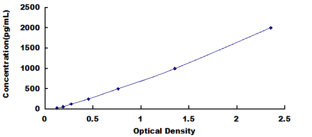
31.25-2000pg/mL. The standard curve concentrations used for the ELISA’s were 2000pg/mL, 1000pg/mL, 500pg/mL, 250pg/mL, 125pg/mL, 62.5pg/mL, 31.2pg/mL.
SENSITIVITY
The minimum detectable dose of AGRN is typically less than 14.6pg/mL.
The sensitivity of this assay, or Lower Limit of Detection (LLD) was defined as the lowest protein concentration that could be differentiated from zero. It was determined by adding two standard deviations to the mean optical density value of twenty zero standard replicates and calculating the corresponding concentration.
SPECIFICITY
This assay has high sensitivity and excellent specificity for detection of AGRN.
No significant cross-reactivity or interference between AGRN and analogues was observed.
You can reference link of the kit as following
https://dldevelop.com/Research-reagent/dl-agrn-hu.html
https://www.dldevelop.com/uploadfile/data/DL-AGRN-Hu.pdf
Introduction
| Item | Standard | Test | |
| Description |
The kit is a sandwich enzyme immunoassay for the in vitro quantitative measurement of AGRN in human serum, plasma, tissue homogenates and other biological fluids. |
Conform | |
| Identification | Colorimetric | Positive | |
| Composition | Traditional ELISA Kit | Ready-to-Use ELISA KIT | Conform |
| Pre-coated, ready to use 96-well strip plate 1 | Pre-coated, ready to use 96-well strip plate 1 | ||
| Plate sealer for 96 wells 2 | Plate sealer for 96 wells 2 | ||
| Standard 2 | Standard 2 | ||
| Diluents buffer 1×45mL | Standard Diluent 1×20mL | ||
| Detection Reagent A 1×120μL | Detection Solution A 1×12mL | ||
| Detection Reagent B 1×120μL | Detection Solution B 1×12mL | ||
| TMB Substrate 1×9mL | TMB Substrate 1×9mL | ||
| Stop Solution 1×6mL | Stop Solution 1×6mL | ||
| Wash Buffer (30 × concentrate) 1×20mL | Wash Buffer (30 × concentrate) 1×20mL | ||
| Instruction manual 1 | Instruction manual 1 | ||
Test principle
The microtiter plate provided in this kit has been pre-coated with an antibody specific to the index. Standards or samples are then added to the appropriate microtiter plate wells with a biotin-conjugated antibody preparation specific to the index. Next, Avidin conjugated to Horseradish Peroxidase (HRP) is added to each microplate well and incubated. After TMB substrate solution is added, only those wells that contain the index, biotin-conjugated antibody and enzyme-conjugated Avidin will exhibit a change in color. The enzyme-substrate reaction is terminated by the addition of sulphuric acid solution and the color change is measured spectrophotometrically at a wavelength of 450nm ± 10nm. The concentration of the index in the samples is then determined by comparing the O.D. of the samples to the standard curve.
Recovery
Matrices listed below were spiked with certain level of recombinant AGRN and the recovery rates were calculated by comparing the measured value to the expected amount of the index in samples.
| Matrix | Recovery range (%) | Average(%) |
| serum(n=5) | 81-93 | 86 |
| EDTA plasma(n=5) | 80-97 | 88 |
| heparin plasma(n=5) | 90-101 | 95 |
Linearity
The linearity of the kit was assayed by testing samples spiked with appropriate concentration of the index and their serial dilutions. The results were demonstrated by the percentage of calculated concentration to the expected.
| Sample | 1:2 | 1:4 | 1:8 | 1:16 |
| serum(n=5) | 82-96% | 83-98% | 81-99% | 93-101% |
| EDTA plasma(n=5) | 88-101% | 86-95% | 90-102% | 80-93% |
| heparin plasma(n=5) | 80-91% | 82-90% | 95-104% | 79-95% |
Precision
Intra-assay Precision (Precision within an assay): 3 samples with low, middle and high level the index were tested 20 times on one plate, respectively.
Inter-assay Precision (Precision between assays): 3 samples with low, middle and high level the index were tested on 3 different plates, 8 replicates in each plate.
CV(%) = SD/meanX100
Intra-Assay: CV<10%
Inter-Assay: CV<12%
Stability
The stability of ELISA kit is determined by the loss rate of activity. The loss rate of this kit is less than 5% within the expiration date under appropriate storage conditions.
Note:
To minimize unnecessary influences on the performance, operation procedures and lab conditions, especially room temperature, air humidity and incubator temperatures should be strictly regulated. It is also strongly suggested that the whole assay is performed by the same experimenter from the beginning to the end.
Assay procedure summary
1. Prepare all reagents, samples and standards;
2. Add 100µL standard or sample to each well. Incubate 2 hours at 37℃;
3. Aspirate and add 100µL prepared Detection Reagent A. Incubate 1 hour at 37℃;
4. Aspirate and wash 3 times;
5. Add 100µL prepared Detection Reagent B. Incubate 1 hour at 37℃;
6. Aspirate and wash 5 times;
7. Add 90µL Substrate Solution. Incubate 15-25 minutes at 37℃;
8. Add 50µL Stop Solution. Read at 450nm immediately.
Order or get a Quote
We will reply you within 24 hours!


