Human Absent In Melanoma 1 (AIM1) ELISA Kit


two product lines: Traditional ELISA Kit and Ready-to-Use ELISA Kit.


Other names:ST4; CRYBG1; Suppression Of Tumorigenicity 4; Beta-Gamma Crystallin Domain Containing 1
Function:May function as suppressor of malignant melanoma. It may exert its effects through interactions with the cytoskeleton.
Sequence:
MEKRSSGRRS GRRRGSQKST DSPGADAELP ESAARDDAVF DDEVAPNAAS
DNASAEKKVK SPRAALDGGV ASAASPESKP SPGTKGQLRG ESDRSKQPPP
ASSPTKRKGR SRALEAVPAP PASGPRAPAK ESPPKRVPDP SPVTKGTAAE
SGEEAARAIP RELPVKSSSL LPEIKPEHKR GPLPNHFNGR AEGGRSRELG
RAAGAPGASD ADGLKPRNHF GVGRSTVTTK VTLPAKPKHV ELNLKTPKNL
DSLGNEHNPF SQPVHKGNTA TKISLFENKR TNSSPRHTDI RGQRNTPASS
KTFVGRAKLN LAKKAKEMEQ PEKKVMPNSP QNGVLVKETA IETKVTVSEE
EILPATRGMN GDSSENQALG PQPNQDDKAD VQTDAGCLSE PVASALIPVK
DHKLLEKEDS EAADSKSLVL ENVTDTAQDI PTTVDTKDLP PTAMPKPQHT
FSDSQSPAES SPGPSLSLSA PAPGDVPKDT CVQSPISSFP CTDLKVSENH
KGCVLPVSRQ NNEKMPLLEL GGETTPPLST ERSPEAVGSE CPSRVLVQVR
SFVLPVESTQ DVSSQVIPES SEVREVQLPT CHSNEPEVVS VASCAPPQEE
VLGNEHSHCT AELAAKSGPQ VIPPASEKTL PIQAQSQGSR TPLMAESSPT
NSPSSGNHLA TPQRPDQTVT NGQDSPASLL NISAGSDDSV FDSSSDMEKF
TEIIKQMDSA VCMPMKRKKA RMPNSPAPHF AMPPIHEDHL EKVFDPKVFT
FGLGKKKESQ PEMSPALHLM QNLDTKSKLR PKRASAEQSV LFKSLHTNTN
GNSEPLVMPE INDKENRDVT NGGIKRSRLE KSALFSSLLS SLPQDKIFSP
SVTSVNTMTT AFSTSQNGSL SQSSVSQPTT EGAPPCGLNK EQSNLLPDNS
LKVFNFNSSS TSHSSLKSPS HMEKYPQKEK TKEDLDSRSN LHLPETKFSE
LSKLKNDDME KANHIESVIK SNLPNCANSD TDFMGLFKSS RYDPSISFSG
MSLSDTMTLR GSVQNKLNPR PGKVVIYSEP DVSEKCIEVF SDIQDCSSWS
LSPVILIKVV RGCWILYEQP NFEGHSIPLE EGELELSGLW GIEDILERHE
EAESDKPVVI GSIRHVVQDY RVSHIDLFTE PEGLGILSSY FDDTEEMQGF
GVMQKTCSMK VHWGTWLIYE EPGFQGVPFI LEPGEYPDLS FWDTEEAYIG
SMRPLKMGGR KVEFPTDPKV VVYEKPFFEG KCVELETGMC SFVMEGGETE
EATGDDHLPF TSVGSMKVLR GIWVAYEKPG FTGHQYLLEE GEYRDWKAWG
GYNGELQSLR PILGDFSNAH MIMYSEKNFG SKGSSIDVLG IVANLKETGY
GVKTQSINVL SGVWVAYENP DFTGEQYILD KGFYTSFEDW GGKNCKISSV
QPICLDSFTG PRRRNQIHLF SEPQFQGHSQ SFEETTSQID DSFSTKSCRV
SGGSWVVYDG ENFTGNQYVL EEGHYPCLSA MGCPPGATFK SLRFIDVEFS
EPTIILFERE DFKGKKIELN AETVNLRSLG FNTQIRSVQV IGGIWVTYEY
GSYRGRQFLL SPAEVPNWYE FSGCRQIGSL RPFVQKRIYF RLRNKATGLF
MSTNGNLEDL KLLRIQVMED VGADDQIWIY QEGCIKCRIA EDCCLTIVGS
LVTSGSKLGL ALDQNADSQF WSLKSDGRIY SKLKPNLVLD IKGGTQYDQN
HIILNTVSKE KFTQVWEAMV LYT
2. Features
INTENDED USE
The kit is a sandwich enzyme immunoassay for the in vitro quantitative measurement of AIM1 in human tissue homogenates, cell lysates and other biological fluids.
DETECTION RANGE
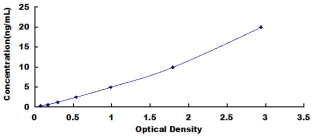
0.312-20ng/mL. The standard curve concentrations used for the ELISA’s were 20ng/mL, 10ng/mL, 5ng/mL, 2.5ng/mL, 1.25ng/mL, 0.625ng/mL, 0.312ng/mL.
SENSITIVITY
The minimum detectable dose of AIM1 is typically less than 0.134ng/mL.
The sensitivity of this assay, or Lower Limit of Detection (LLD) was defined as the lowest protein concentration that could be differentiated from zero. It was determined by adding two standard deviations to the mean optical density value of twenty zero standard replicates and calculating the corresponding concentration.
SPECIFICITY
This assay has high sensitivity and excellent specificity for detection of AIM1.
No significant cross-reactivity or interference between AIM1 and analogues was observed.
Note: Limited by current skills and knowledge, it is impossible to perform all possible cross-reactivity detection tests between AIM1 and all analogues, therefore, cross reactivity may still exist.
IMPORTANT NOTES
1. Limited by the current conditions and scientific technology, it is impossible to conduct comprehensive identification and analysis tests on the raw materials provided by suppliers. As a result, it is possible there are some qualitative and/or technical risks.
2. The final experimental results will be closely related to the validity of the products, operation skills of the end users and the experimental environments. Please make sure that sufficient samples are available to obtain accurate results.
3. Kits from different batches may be a little different in detection range, sensitivity and color developing time. Please perform the experiment exactly according to the instruction manual included in your kit. Electronic ones on our website are for reference only.
4. Do not mix or substitute reagents from one kit lot to another. Use only the reagents supplied by manufacturer.
5. Protect all reagents from strong light during storage and incubation. All bottle caps of reagents should be closed tightly to prevent evaporation of liquids and contamination by microorganisms.
6. There may be a foggy substance in the wells when the plate is opened at the first time. It will not have any effect on the final assay results. Do not remove microtiter plate from the storage bag until needed.
7. Incorrect procedures during reagent preparation and loading, as well as incorrect parameter setting for the plate reader may lead to incorrect results. A microplate plate reader with a bandwidth of 10nm or less and an optical density range of 0-3 O.D. or greater at 450 ± 10nm wavelength is acceptable for use in absorbance measurement. Please read the instruction carefully and adjust the instrument prior to the experiment.
8. Even the same experimenter may get different results from two separate experiments. In order to get better reproducible results, the operation of every step in the assay should be controlled. Furthermore, a preliminary experiment before the general assay for each batch is recommended.
9. Each kit has undergone several rigorous quality control tests. However, results from end users might be inconsistent with our in-house data due to some unexpected transportation conditions or different lab equipment. Intra-assay variance among kits from different batches could arise from the above factors as well.
10. Kits from different manufacturers with the same item might produce different results, since we have not compared our products with other manufacturers.
11. The standard in this kit, as well as the antigens used in antibody preparation are typically recombinant proteins. Differently expressed sequences, expression systems, and/or purification methods can be used in the preparation of recombinant proteins. There is also the possibility of differences in the screening technique of antibodies and antibody pairs in our kits. As a result, we cannot guarantee that our kit will be able to detect recombinant proteins produced by other companies. We do NOT recommend using our ELISA kits for the detection of other recombinant proteins.
12. Validity period: 12 months.
13. The instruction manual also works with the 48T kit, but all reagents in the 48T kit are reduced by half.
You can reference link of the kit as following
https://dldevelop.com/Research-reagent/dl-aim1-hu.html
https://www.dldevelop.com/uploadfile/data/DL-AIM1-Hu.pdf
Introduction
| Item | Standard | Test | |
| Description |
The kit is a sandwich enzyme immunoassay for the in vitro quantitative measurement of AIM1 in human tissue homogenates, cell lysates or other biological fluids. |
Conform | |
| Identification | Colorimetric | Positive | |
| Composition | Traditional ELISA Kit | Ready-to-Use ELISA KIT | Conform |
| Pre-coated, ready to use 96-well strip plate 1 | Pre-coated, ready to use 96-well strip plate 1 | ||
| Plate sealer for 96 wells 2 | Plate sealer for 96 wells 2 | ||
| Standard 2 | Standard 2 | ||
| Diluents buffer 1×45mL | Standard Diluent 1×20mL | ||
| Detection Reagent A 1×120μL | Detection Solution A 1×12mL | ||
| Detection Reagent B 1×120μL | Detection Solution B 1×12mL | ||
| TMB Substrate 1×9mL | TMB Substrate 1×9mL | ||
| Stop Solution 1×6mL | Stop Solution 1×6mL | ||
| Wash Buffer (30 × concentrate) 1×20mL | Wash Buffer (30 × concentrate) 1×20mL | ||
| Instruction manual 1 | Instruction manual 1 | ||
Test principle
The microtiter plate provided in this kit has been pre-coated with an antibody specific to the index. Standards or samples are then added to the appropriate microtiter plate wells with a biotin-conjugated antibody preparation specific to the index. Next, Avidin conjugated to Horseradish Peroxidase (HRP) is added to each microplate well and incubated. After TMB substrate solution is added, only those wells that contain the index, biotin-conjugated antibody and enzyme-conjugated Avidin will exhibit a change in color. The enzyme-substrate reaction is terminated by the addition of sulphuric acid solution and the color change is measured spectrophotometrically at a wavelength of 450nm ± 10nm. The concentration of the index in the samples is then determined by comparing the O.D. of the samples to the standard curve.
Recovery
Matrices listed below were spiked with certain level of recombinant AIM1 and the recovery rates were calculated by comparing the measured value to the expected amount of the index in samples.
| Matrix | Recovery range (%) | Average(%) |
| serum(n=5) | 81-93 | 86 |
| EDTA plasma(n=5) | 80-97 | 88 |
| heparin plasma(n=5) | 90-101 | 95 |
Linearity
The linearity of the kit was assayed by testing samples spiked with appropriate concentration of the index and their serial dilutions. The results were demonstrated by the percentage of calculated concentration to the expected.
| Sample | 1:2 | 1:4 | 1:8 | 1:16 |
| serum(n=5) | 82-96% | 83-98% | 81-99% | 93-101% |
| EDTA plasma(n=5) | 88-101% | 86-95% | 90-102% | 80-93% |
| heparin plasma(n=5) | 80-91% | 82-90% | 95-104% | 79-95% |
Precision
Intra-assay Precision (Precision within an assay): 3 samples with low, middle and high level the index were tested 20 times on one plate, respectively.
Inter-assay Precision (Precision between assays): 3 samples with low, middle and high level the index were tested on 3 different plates, 8 replicates in each plate.
CV(%) = SD/meanX100
Intra-Assay: CV<10%
Inter-Assay: CV<12%
Stability
The stability of ELISA kit is determined by the loss rate of activity. The loss rate of this kit is less than 5% within the expiration date under appropriate storage conditions.
Note:
To minimize unnecessary influences on the performance, operation procedures and lab conditions, especially room temperature, air humidity and incubator temperatures should be strictly regulated. It is also strongly suggested that the whole assay is performed by the same experimenter from the beginning to the end.
Assay procedure summary
1. Prepare all reagents, samples and standards;
2. Add 100µL standard or sample to each well. Incubate 2 hours at 37℃;
3. Aspirate and add 100µL prepared Detection Reagent A. Incubate 1 hour at 37℃;
4. Aspirate and wash 3 times;
5. Add 100µL prepared Detection Reagent B. Incubate 1 hour at 37℃;
6. Aspirate and wash 5 times;
7. Add 90µL Substrate Solution. Incubate 15-25 minutes at 37℃;
8. Add 50µL Stop Solution. Read at 450nm immediately.
Order or get a Quote
We will reply you within 24 hours!


