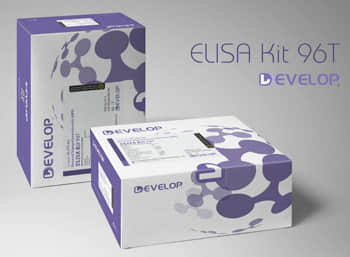
Human Adaptor Related Protein Complex 2 Mu 1 (AP2m1) ELISA Kit


two product lines: Traditional ELISA Kit and Ready-to-Use ELISA Kit.


Other names:AP50; CLAPM1; Clathrin-Associated/Assembly/Adaptor Protein,Medium 1; Plasma Membrane Adaptor AP-2 50kDA Protein; Clathrin Coat Adaptor Protein AP50
Function: Component of the adaptor protein complex 2 (AP-2). Adaptor protein complexes function in protein transport via transport vesicles in different membrane traffic pathways. Adaptor protein complexes are vesicle coat components and appear to be involved in cargo selection and vesicle formation. AP-2 is involved in clathrin-dependent endocytosis in which cargo proteins are incorporated into vesicles surrounded by clathrin (clathrin-coated vesicles, CCVs) which are destined for fusion with the early endosome. The clathrin lattice serves as a mechanical scaffold but is itself unable to bind directly to membrane components. Clathrin-associated adaptor protein (AP) complexes which can bind directly to both the clathrin lattice and to the lipid and protein components of membranes are considered to be the major clathrin adaptors contributing the CCV formation. AP-2 also serves as a cargo receptor to selectively sort the membrane proteins involved in receptor-mediated endocytosis. AP-2 seems to play a role in the recycling of synaptic vesicle membranes from the presynaptic surface. AP-2 recognizes Y-X-X-[FILMV] (Y-X-X-Phi) and [ED]-X-X-X-L-[LI] endocytosis signal motifs within the cytosolic tails of transmembrane cargo molecules. AP-2 may also play a role in maintaining normal post-endocytic trafficking through the ARF6-regulated, non-clathrin pathway. The AP-2 mu subunit binds to transmembrane cargo proteins; it recognizes the Y-X-X-Phi motifs. The surface region interacting with to the Y-X-X-Phi motif is inaccessible in cytosolic AP-2, but becomes accessible through a conformational change following phosphorylation of AP-2 mu subunit at 'Tyr-156' in membrane-associated AP-2. The membrane-specific phosphorylation event appears to involve assembled clathrin which activates the AP-2 mu kinase AAK1 (By similarity). Plays a role in endocytosis of frizzled family members upon Wnt signaling (By similarity).
Sequence:
 50 MIGGLFIYNH KGEVLISRVY RDDIGRNAVD AFRVNVIHAR QQVRSPVTNI
100 ARTSFFHVKR SNIWLAAVTK QNVNAAMVFE FLYKMCDVMA AYFGKISEEN
150 IKNNFVLIYE LLDEILDFGY PQNSETGALK TFITQQGIKS QHQTKEEQSQ
200 ITSQVTGQIG WRREGIKYRR NELFLDVLES VNLLMSPQGQ VLSAHVSGRV
250 VMKSYLSGMP ECKFGMNDKI VIEKQGKGTA DETSKSGKQS IAIDDCTFHQ
300 CVRLSKFDSE RSISFIPPDG EFELMRYRTT KDIILPFRVI PLVREVGRTK
350 LEVKVVIKSN FKPSLLAQKI EVRIPTPLNT SGVQVICMKG KAKYKASENA
400 IVWKIKRMAG MKESQISAEI ELLPTNDKKK WARPPISMNF EVPFAPSGLK
430 VRYLKVFEPK LNYSDHDVIK WVRYIGRSGI YETRC
INTENDED USE
The kit is a sandwich enzyme immunoassay for the in vitro quantitative measurement of AP2m1 in human tissue homogenates or other biological fluids.
DETECTION RANGE
46.875-3000pg/mL. The standard curve concentrations used for the ELISA’s were 3000pg/mL, 1500pg/mL, 750pg/mL, 375pg/mL, 187pg/mL, 93.7pg/mL, 46.8pg/mL.
SENSITIVITY
The minimum detectable dose of AMH is typically less than 23.2pg/mL.
The sensitivity of this assay, or Lower Limit of Detection (LLD) was defined as the lowest protein concentration that could be differentiated from zero. It was determined by adding two standard deviations to the mean optical density value of twenty zero standard replicates and calculating the corresponding concentration.
SPECIFICITY
This assay has high sensitivity and excellent specificity for detection of AMH.
No significant cross-reactivity or interference between AMH and analogues was observed.
You can reference link of the kit as following
https://dldevelop.com/Research-reagent/dl-ap2m1-hu.html
https://www.dldevelop.com/uploadfile/data/DL-AP2m1-Hu.pdf
Introduction
| Item | Standard | Test | |
| Description |
The kit is a sandwich enzyme immunoassay for the in vitro quantitative measurement of AP2m1 in human tissue homogenates or other biological fluids. |
Conform | |
| Identification | Colorimetric | Positive | |
| Composition | Traditional ELISA Kit | Ready-to-Use ELISA KIT | Conform |
| Pre-coated, ready to use 96-well strip plate 1 | Pre-coated, ready to use 96-well strip plate 1 | ||
| Plate sealer for 96 wells 2 | Plate sealer for 96 wells 2 | ||
| Standard 2 | Standard 2 | ||
| Diluents buffer 1×45mL | Standard Diluent 1×20mL | ||
| Detection Reagent A 1×120μL | Detection Solution A 1×12mL | ||
| Detection Reagent B 1×120μL | Detection Solution B 1×12mL | ||
| TMB Substrate 1×9mL | TMB Substrate 1×9mL | ||
| Stop Solution 1×6mL | Stop Solution 1×6mL | ||
| Wash Buffer (30 × concentrate) 1×20mL | Wash Buffer (30 × concentrate) 1×20mL | ||
| Instruction manual 1 | Instruction manual 1 | ||
Test principle
The microtiter plate provided in this kit has been pre-coated with an antibody specific to the index. Standards or samples are then added to the appropriate microtiter plate wells with a biotin-conjugated antibody preparation specific to the index. Next, Avidin conjugated to Horseradish Peroxidase (HRP) is added to each microplate well and incubated. After TMB substrate solution is added, only those wells that contain the index, biotin-conjugated antibody and enzyme-conjugated Avidin will exhibit a change in color. The enzyme-substrate reaction is terminated by the addition of sulphuric acid solution and the color change is measured spectrophotometrically at a wavelength of 450nm ± 10nm. The concentration of the index in the samples is then determined by comparing the O.D. of the samples to the standard curve.
Recovery
Matrices listed below were spiked with certain level of recombinant AP2m1 and the recovery rates were calculated by comparing the measured value to the expected amount of the index in samples.
| Matrix | Recovery range (%) | Average(%) |
| serum(n=5) | 81-93 | 86 |
| EDTA plasma(n=5) | 80-97 | 88 |
| heparin plasma(n=5) | 90-101 | 95 |
Linearity
The linearity of the kit was assayed by testing samples spiked with appropriate concentration of the index and their serial dilutions. The results were demonstrated by the percentage of calculated concentration to the expected.
| Sample | 1:2 | 1:4 | 1:8 | 1:16 |
| serum(n=5) | 82-96% | 83-98% | 81-99% | 93-101% |
| EDTA plasma(n=5) | 88-101% | 86-95% | 90-102% | 80-93% |
| heparin plasma(n=5) | 80-91% | 82-90% | 95-104% | 79-95% |
Precision
Intra-assay Precision (Precision within an assay): 3 samples with low, middle and high level the index were tested 20 times on one plate, respectively.
Inter-assay Precision (Precision between assays): 3 samples with low, middle and high level the index were tested on 3 different plates, 8 replicates in each plate.
CV(%) = SD/meanX100
Intra-Assay: CV<10%
Inter-Assay: CV<12%
Stability
The stability of ELISA kit is determined by the loss rate of activity. The loss rate of this kit is less than 5% within the expiration date under appropriate storage conditions.
Note:
To minimize unnecessary influences on the performance, operation procedures and lab conditions, especially room temperature, air humidity and incubator temperatures should be strictly regulated. It is also strongly suggested that the whole assay is performed by the same experimenter from the beginning to the end.
Assay procedure summary
1. Prepare all reagents, samples and standards;
2. Add 100µL standard or sample to each well. Incubate 2 hours at 37℃;
3. Aspirate and add 100µL prepared Detection Reagent A. Incubate 1 hour at 37℃;
4. Aspirate and wash 3 times;
5. Add 100µL prepared Detection Reagent B. Incubate 1 hour at 37℃;
6. Aspirate and wash 5 times;
7. Add 90µL Substrate Solution. Incubate 15-25 minutes at 37℃;
8. Add 50µL Stop Solution. Read at 450nm immediately.
Order or get a Quote
We will reply you within 24 hours!


