Mouse Fibroblast Growth Factor Receptor 2 (FGFR2) ELISA Kit


two product lines: Traditional ELISA Kit and Ready-to-Use ELISA Kit.


Other names:Keratinocyte growth factor receptor(KGFR)
Function: Tyrosine-protein kinase that acts as cell-surface receptor for fibroblast growth factors and plays an essential role in the regulation of cell proliferation, differentiation, migration and apoptosis, and in the regulation of embryonic development. Required for normal embryonic patterning, trophoblast function, limb bud development, lung morphogenesis, osteogenesis and skin development. Plays an essential role in the regulation of osteoblast differentiation, proliferation and apoptosis, and is required for normal skeleton development. Promotes cell proliferation in keratinocytes and imature osteoblasts, but promotes apoptosis in differentiated osteoblasts. Phosphorylates PLCG1, FRS2A and PAK4. Ligand binding leads to the activation of several signaling cascades. Activation of PLCG1 leads to the production of the cellular signaling molecules diacylglycerol and inositol-1,4,5-trisphosphate. Phosphorylation of FRS2A triggers recruitment of GRB2, GAB1, PIK3R1 and SOS1, and mediates activation of RAS, MAPK1/ERK2, MAPK3/ERK1 and the MAP kinase signaling pathway, as well as of the AKT1 signaling pathway. FGFR2 signaling is down-regulated by ubiquitination, internalization and degradation. Mutations that lead to constitutive kinase activation or impair normal FGFR2 maturation, internalization and degradation lead to aberrant signaling. Over-expressed FGFR2 promotes activation of STAT1.
Sequence:
50 MVSWGRFICL VLVTMATLSL ARPSFSLVED TTLEPEEPPT KYQISQPEAY
100 VVAPGESLEL QCMLKDAAVI SWTKDGVHLG PNNRTVLIGE YLQIKGAT
150 DSGLYACTAA RTVDSETWIF MVNVTDAISS GDDEDDTDSS EDVVSENRSN
200 QRAPYWTNTE KMEKRLHACP AANTVKFRCP AGGNPTSTMR WLKNGKEFKQ
250 EHRIGGYKVR NQHWSLIMES VVPSDKGNYT CLVENEYGSI NHTYHLDVVE
300 RSPHRPILQA GLPANASTVV GGDVEFVCKV YSDAQPHIQW IKHVEKNGSK
350 NGPDGLPYLK VLKAAGVNTT DKEIEVLYIR NVTFEDAGEY TCLAGNSIGI
400 SFHSAWLTVL PAPVREKEIT ASPDYLEIAI YCIGVFLIAC MVVTVIFCRM
450 KTTTKKPDFS SQPAVHKLTK RIPLRRQVTV SAESSSSMNS NTPLVRITTR
500 LSSTADTPML AGVSEYELPE DPKWEFPRDK LTLGKPLGEG CFGQVVMAEA
550 VGIDKDKPKE AVTVAVKMLK DDATEKDLSD LVSEMEMMKM IGKHKNIINL
600 LGACTQDGPL YVIVEYASKG NLREYLRARR PPGMEYSYDI NRVPEEQMTF
650 KDLVSCTYQL ARGMEYLASQ KCIHRDLAAR NVLVTENNVM KIADFGLARD
700 INNIDYYKKT TNGRLPVKWM APEALFDRVY THQSDVWSFG VLMWEIFTLG
750 GSPYPGIPVE ELFKLLKEGH RMDKPTNCTN ELYMMMRDCW HAVPSQRPTF
800 KQLVEDLDRI LTLTTNEEYL DLTQPLEQYS PSYPDTSSSC SSGDDSVFSP
820 DPMPYEPCLP QYPHINGSVK T
INTENDED USE
The kit is a sandwich enzyme immunoassay for the in vitro quantitative measurement of FGFR2 in mouse serum, plasma, tissue homogenates or other biological fluids.
DETECTION RANGE
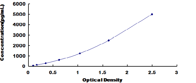
78.125-5000pg/mL. The standard curve concentrations used for the ELISA’s were 5000pg/mL, 2500pg/mL, 1250pg/mL, 625pg/mL, 312pg/mL, 156pg/mL, 78.1pg/mL.
SENSITIVITY
The minimum detectable dose of FGFR2 is typically less than 30.1pg/mL.
The sensitivity of this assay, or Lower Limit of Detection (LLD) was defined as the lowest protein concentration that could be differentiated from zero. It was determined by adding two standard deviations to the mean optical density value of twenty zero standard replicates and calculating the corresponding concentration.
SPECIFICITY
This assay has high sensitivity and excellent specificity for detection of FGFR2.
No significant cross-reactivity or interference between FGFR2 and analogues was observed.
You can reference link of the kit as following
https://dldevelop.com/Research-reagent/dl-fgfr2-mu.html
https://www.dldevelop.com/uploadfile/data/DL-FGFR2-Mu.pdf
Introduction
| Item | Standard | Test | |
| Description |
The kit is a sandwich enzyme immunoassay for the in vitro quantitative measurement of FGFR2 in mouse serum, plasma, tissue homogenates or other biological fluids. |
Conform | |
| Identification | Colorimetric | Positive | |
| Composition | Traditional ELISA Kit | Ready-to-Use ELISA KIT | Conform |
| Pre-coated, ready to use 96-well strip plate 1 | Pre-coated, ready to use 96-well strip plate 1 | ||
| Plate sealer for 96 wells 2 | Plate sealer for 96 wells 2 | ||
| Standard 2 | Standard 2 | ||
| Diluents buffer 1×45mL | Standard Diluent 1×20mL | ||
| Detection Reagent A 1×120μL | Detection Solution A 1×12mL | ||
| Detection Reagent B 1×120μL | Detection Solution B 1×12mL | ||
| TMB Substrate 1×9mL | TMB Substrate 1×9mL | ||
| Stop Solution 1×6mL | Stop Solution 1×6mL | ||
| Wash Buffer (30 × concentrate) 1×20mL | Wash Buffer (30 × concentrate) 1×20mL | ||
| Instruction manual 1 | Instruction manual 1 | ||
Test principle
The microtiter plate provided in this kit has been pre-coated with an antibody specific to the index. Standards or samples are then added to the appropriate microtiter plate wells with a biotin-conjugated antibody preparation specific to the index. Next, Avidin conjugated to Horseradish Peroxidase (HRP) is added to each microplate well and incubated. After TMB substrate solution is added, only those wells that contain the index, biotin-conjugated antibody and enzyme-conjugated Avidin will exhibit a change in color. The enzyme-substrate reaction is terminated by the addition of sulphuric acid solution and the color change is measured spectrophotometrically at a wavelength of 450nm ± 10nm. The concentration of the index in the samples is then determined by comparing the O.D. of the samples to the standard curve.
Recovery
Matrices listed below were spiked with certain level of recombinant FGFR2 and the recovery rates were calculated by comparing the measured value to the expected amount of the index in samples.
| Matrix | Recovery range (%) | Average(%) |
| serum(n=5) | 81-93 | 86 |
| EDTA plasma(n=5) | 80-97 | 88 |
| heparin plasma(n=5) | 90-101 | 95 |
Linearity
The linearity of the kit was assayed by testing samples spiked with appropriate concentration of the index and their serial dilutions. The results were demonstrated by the percentage of calculated concentration to the expected.
| Sample | 1:2 | 1:4 | 1:8 | 1:16 |
| serum(n=5) | 82-96% | 83-98% | 81-99% | 93-101% |
| EDTA plasma(n=5) | 88-101% | 86-95% | 90-102% | 80-93% |
| heparin plasma(n=5) | 80-91% | 82-90% | 95-104% | 79-95% |
Precision
Intra-assay Precision (Precision within an assay): 3 samples with low, middle and high level the index were tested 20 times on one plate, respectively.
Inter-assay Precision (Precision between assays): 3 samples with low, middle and high level the index were tested on 3 different plates, 8 replicates in each plate.
CV(%) = SD/meanX100
Intra-Assay: CV<10%
Inter-Assay: CV<12%
Stability
The stability of ELISA kit is determined by the loss rate of activity. The loss rate of this kit is less than 5% within the expiration date under appropriate storage conditions.
Note:
To minimize unnecessary influences on the performance, operation procedures and lab conditions, especially room temperature, air humidity and incubator temperatures should be strictly regulated. It is also strongly suggested that the whole assay is performed by the same experimenter from the beginning to the end.
Assay procedure summary
1. Prepare all reagents, samples and standards;
2. Add 100µL standard or sample to each well. Incubate 2 hours at 37℃;
3. Aspirate and add 100µL prepared Detection Reagent A. Incubate 1 hour at 37℃;
4. Aspirate and wash 3 times;
5. Add 100µL prepared Detection Reagent B. Incubate 1 hour at 37℃;
6. Aspirate and wash 5 times;
7. Add 90µL Substrate Solution. Incubate 15-25 minutes at 37℃;
8. Add 50µL Stop Solution. Read at 450nm immediately.
Order or get a Quote
We will reply you within 24 hours!


