


![]() 3 times QC before delivery.
3 times QC before delivery.
![]() 12-month shelf life.
12-month shelf life.
![]() Strong technical support
Strong technical support
![]() Prompt response
Prompt response
After-sales service guarantee.

| > Cytokine |
| > Signal transduction |
| > Enzyme & Kinase |
| > Metabolic pathway |
| > Tumor immunity |
| > Infection immunity |
| > CD& Adhesion molecule |
| > Neuro science |
| > Developmental science |

Mouse High Mobility Group Protein 1 (HMG1) ELISA Kit

two product lines: Traditional ELISA Kit and Ready-to-Use ELISA Kit.
| Traditional ELISA Kit | Ready-to-Use ELISA KIT | |
| Product name: | Mouse High Mobility Group Protein 1 (HMG1) ELISA Kit | |
| Method: | Sandwich | |
| Synonyms: | HMGB1; HMG3; SBP1; Sulfoglucuronyl Carbohydrate Binding Protein; Amphoterin; High Mobility Group Box 1 Protein |
|
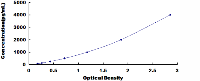
| Detection range: | 62.5-4,000pg/mL | |
| Target Protein: | HMG1 | |
| Size: | 96T/48T | |
| Quality guarantee period: | for 12 months, 16 months | |
| Catalog number: | DL-HMG1-Mu (traditional) | (ready-to-use) |
| Assay length | 1-4.5Hours | 1-3.5Hours |
| Advantages: |
|
|
| Instruction Manual |
|
|
Overview
Other names: HMGB1; HMG3; SBP1; Sulfoglucuronyl Carbohydrate Binding Protein; Amphoterin; High Mobility Group Box 1 Protein
Function: Multifunctional redox sensitive protein with various roles in different cellular compartments. In the nucleus is one of the major chromatin-associated non-histone proteins and acts as a DNA chaperone involved in replication, transcription, chromatin remodeling, V(D)J recombination, DNA repair and genome stability. Proposed to be an universal biosensor for nucleic acids. Promotes host inflammatory response to sterile and infectious signals and is involved in the coordination and integration of innate and adaptive immune responses. In the cytoplasm functions as sensor and/or chaperone for immunogenic nucleic acids implicating the activation of TLR9-mediated immune responses, and mediates autophagy. Acts as danger associated molecular pattern (DAMP) molecule that amplifies immune responses during tissue injury. Released to the extracellular environment can bind DNA, nucleosomes, IL-1 beta, CXCL12, AGER isoform 2/sRAGE, lipopolysaccharide (LPS) and lipoteichoic acid (LTA), and activates cells through engagement of multiple surface receptors. In the extracellular compartment fully reduced HMGB1 (released by necrosis) acts as a chemokine, disulfide HMGB1 (actively secreted) as a cytokine, and sulfonyl HMGB1 (released from apoptotic cells) promotes immunological tolerance (PubMed:23519706, PubMed:23446148, PubMed:23994764, PubMed:25048472). Has proangiogenic activity (PubMed:16365390). May be involved in platelet activation. Binds to phosphatidylserine and phosphatidylethanolamide. Bound to RAGE mediates signaling for neuronal outgrowth. May play a role in accumulation of expanded polyglutamine (polyQ) proteins (By similarity).By similarity4 Publications1 Publication Nuclear functions are attributed to fully reduced HGMB1. Associates with chromatin and binds DNA with a preference to non-canonical DNA structures such as single-stranded DNA, DNA-containing cruciforms or bent structures, supercoiled DNA and ZDNA. Can bent DNA and enhance DNA flexibility by looping thus providing a mechanism to promote activities on various gene promoters by enhancing transcription factor binding and/or bringing distant regulatory sequences into close proximity. May be involved in nucleotide excision repair (NER), mismatch repair (MMR) and base excision repair (BER) pathways, and double strand break repair such as non-homologous end joining (NHEJ) (PubMed:17803946, PubMed:18650382). Involved in V(D)J recombination by acting as a cofactor of the RAG complex: acts by stimulating cleavage and RAG protein binding at the 23 bp spacer of conserved recombination signal sequences (RSS). In vitro can displace histone H1 from highly bent DNA. Can restructure the canonical nucleosome leading to relaxation of structural constraints for transcription factor-binding (By similarity). Enhances binding of sterol regulatory element-binding proteins (SREBPs) such as SREBF1 to their cognate DNA sequences and increases their transcriptional activities (PubMed:16040616). Facilitates binding of TP53 to DNA (By similarity). Proposed to be involved in mitochondrial quality control and autophagy in a transcription-dependent fashion implicating HSPB1; however, this function has been questioned (PubMed:21641551, PubMed:24606906). Can modulate the activity of the telomerase complex and may be involved in telomere maintenance (PubMed:22544226).By similarity6 Publications
In the cytoplasm proposed to dissociate the BECN1:BCL2 complex via competitive interaction with BECN1 leading to autophagy activation (PubMed:21395369). Can protect BECN1 and ATG5 from calpain-mediated cleavage and thus proposed to control their proautophagic and proapoptotic functions and to regulate the extent and severity of inflammation-associated cellular injury (PubMed:25642769). In myeloid cells has a protective role against endotoxemia and bacterial infection by promoting autophagy (PubMed:24302768). Involved in endosomal translocation and activation of TLR9 in response to CpG-DNA in macrophages (PubMed:17548579).By similarity5 Publications In the extracellular compartment (following either active secretion or passive release) involved in regulation of the inflammatory response. Fully reduced HGMB1 (which subsequently gets oxidized after release) in association with CXCL12 mediates the recruitment of inflammatory cells during the initial phase of tissue injury; the CXCL12:HMGB1 complex triggers CXCR4 homodimerization (PubMed:22370717). Induces the migration of monocyte-derived immature dendritic cells and seems to regulate adhesive and migratory functions of neutrophils implicating AGER/RAGE and ITGAM (PubMed:17268551). Can bind to various types of DNA and RNA including microbial unmethylated CpG-DNA to enhance the innate immune response to nucleic acids. Proposed to act in promiscuous DNA/RNA sensing which cooperates with subsequent discriminative sensing by specific pattern recognition receptors (PubMed:19890330). Promotes extracellular DNA-induced AIM2 inflammasome activation implicating AGER/RAGE.
Disulfide HMGB1 binds to transmembrane receptors, such as AGER/RAGE, TLR2, TLR4 and probably TREM1, thus activating their signal transduction pathways (PubMed:17568691, PubMed:19264983, PubMed:21419643). Mediates the release of cytokines/chemokines such as TNF, IL-1, IL-6, IL-8, CCL2, CCL3, CCL4 and CXCL10 (PubMed:12110890, PubMed:17548579). Promotes secretion of interferon-gamma by macrophage-stimulated natural killer (NK) cells in concert with other cytokines like IL-2 or IL-12. TLR4 is proposed to be the primary receptor promoting macrophage activation and signaling through TLR4 seems to implicate LY96/MD-2. In bacterial LPS- or LTA-mediated inflammatory responses binds to the endotoxins and transfers them to CD14 for signaling to the respective TLR4:LY96 and TLR2 complexes (By similarity). Contributes to tumor proliferation by association with ACER/RAGE (By similarity). Can bind to IL1-beta and signals through the IL1R1:IL1RAP receptor complex (By similarity). Binding to class A CpG activates cytokine production in plasmacytoid dendritic cells implicating TLR9, MYD88 and AGER/RAGE and can activate autoreactive B cells. Via HMGB1-containing chromatin immune complexes may also promote B cell responses to endogenous TLR9 ligands through a B-cell receptor (BCR)-dependent and ACER/RAGE-independent mechanism (By similarity). Inhibits phagocytosis of apoptotic cells by macrophages; the function is dependent on poly-ADP-ribosylation and involves binding to phosphatidylserine on the cell surface of apoptotic cells (PubMed:22204001, PubMed:18768881). In adaptive immunity may be involved in enhancing immunity through activation of effector T cells and suppression of regulatory T (TReg) cells (PubMed:21419643). In contrast, without implicating effector or regulatory T cells, required for tumor infiltration and activation of T cells expressing the lymphotoxin LTA:LTB heterotrimer thus promoting tumor malignant progression (PubMed:23108142). Also reported to limit proliferation of T cells (By similarity). Released HMGB1:nucleosome complexes formed during apoptosis can signal through TLR2 to induce cytokine production (By similarity). Involved in induction of immunological tolerance by apoptotic cells; its pro-inflammatory activities when released by apoptotic cells are neutralized by reactive oxygen species (ROS)-dependent oxidation specifically on Cys-106 (By similarity). During macrophage activation by activated lymphocyte-derived self apoptotic DNA (ALD-DNA) promotes recruitment of ALD-DNA to endosomes (PubMed:25660970).
Sequence:
MGKGDPKKPR GKMSSYAFFV QTCREEHKKK HPDASVNFSE FSKKCSERWK
TMSAKEKGKF EDMAKADKAR YEREMKTYIP PKGETKKKFK DPNAPKRPPS
AFFLFCSEYR PKIKGEHPGL SIGDVAKKLG EMWNNTAADD KQPYEKKAAK
LKEKYEKDIA AYRAKGKPDA AKKGVVKAEK SKKKKEEEDD EEDEEDEEEE
EEEEDEDEEE DDDDE
KYEMVPNLIT QHCACI
Features
DL-HMG1-Mu
Mouse High Mobility Group Protein 1 (HMG1) ELISA Kit
INTENDED USE
The kit is a sandwich enzyme immunoassay for in vitro quantitative measurement of HMG1 in mouse serum, plasma and other biological fluids.
SENSITIVITY
The minimum detectable dose of this mouse HMG1 is typically less than 0.12ng/mL
The sensitivity of this assay, or Lower Limit of Detection (LLD) was defined as the lowest protein concentration that could be differentiated from zero. It was determined by adding two standard deviations to the mean optical density value of twenty zero standard replicates and calculating the corresponding concentration.
SPECIFICITY
This assay has high sensitivity and excellent specificity for detection of mouse HMG1.
No significant cross-reactivity or interference between mouse HMG1 and analogues was observed. Note: Limited by current skills and knowledge, it is impossible for us to complete the cross- reactivity detection between mouse HMG1 and all the analogues, therefore, cross reaction may still exist.
We have this HMG1 Elisa kit for mouse mouse rat rabbit pig dog monkey and Bovine
You can reference link of the kit as following
Introduction
| Item | Standard | Test | |
| Description |
The kit is a sandwich enzyme immunoassay for the in vitro quantitative measurement of HMG1 in mouse serum, plasma and other biological fluids. |
Conform | |
| Identification | Colorimetric | Positive | |
| Composition | Traditional ELISA Kit | Ready-to-Use ELISA KIT | Conform |
| Pre-coated, ready to use 96-well strip plate 1 | Pre-coated, ready to use 96-well strip plate 1 | ||
| Plate sealer for 96 wells 2 | Plate sealer for 96 wells 2 | ||
| Standard 2 | Standard 2 | ||
| Diluents buffer 1×45mL | Standard Diluent 1×20mL | ||
| Detection Reagent A 1×120μL | Detection Solution A 1×12mL | ||
| Detection Reagent B 1×120μL | Detection Solution B 1×12mL | ||
| TMB Substrate 1×9mL | TMB Substrate 1×9mL | ||
| Stop Solution 1×6mL | Stop Solution 1×6mL | ||
| Wash Buffer (30 × concentrate) 1×20mL | Wash Buffer (30 × concentrate) 1×20mL | ||
| Instruction manual 1 | Instruction manual 1 | ||
Test principle
The microtiter plate provided in this kit has been pre-coated with an antibody specific to the index. Standards or samples are then added to the appropriate microtiter plate wells with a biotin-conjugated antibody preparation specific to the index. Next, Avidin conjugated to Horseradish Peroxidase (HRP) is added to each microplate well and incubated. After TMB substrate solution is added, only those wells that contain the index, biotin-conjugated antibody and enzyme-conjugated Avidin will exhibit a change in color. The enzyme-substrate reaction is terminated by the addition of sulphuric acid solution and the color change is measured spectrophotometrically at a wavelength of 450nm ± 10nm. The concentration of the index in the samples is then determined by comparing the O.D. of the samples to the standard curve.
Recovery
Matrices listed below were spiked with certain level of recombinant HMG1 and the recovery rates were calculated by comparing the measured value to the expected amount of the index in samples.
| Matrix | Recovery range (%) | Average(%) |
| serum(n=5) | 81-93 | 86 |
| EDTA plasma(n=5) | 80-97 | 88 |
| heparin plasma(n=5) | 90-101 | 95 |
Linearity
The linearity of the kit was assayed by testing samples spiked with appropriate concentration of the index and their serial dilutions. The results were demonstrated by the percentage of calculated concentration to the expected.
| Sample | 1:2 | 1:4 | 1:8 | 1:16 |
| serum(n=5) | 82-96% | 83-98% | 81-99% | 93-101% |
| EDTA plasma(n=5) | 88-101% | 86-95% | 90-102% | 80-93% |
| heparin plasma(n=5) | 80-91% | 82-90% | 95-104% | 79-95% |
Precision
Intra-assay Precision (Precision within an assay): 3 samples with low, middle and high level the index were tested 20 times on one plate, respectively.
Inter-assay Precision (Precision between assays): 3 samples with low, middle and high level the index were tested on 3 different plates, 8 replicates in each plate.
CV(%) = SD/meanX100
Intra-Assay: CV<10%
Inter-Assay: CV<12%
Stability
The stability of ELISA kit is determined by the loss rate of activity. The loss rate of this kit is less than 5% within the expiration date under appropriate storage conditions.
Note:
To minimize unnecessary influences on the performance, operation procedures and lab conditions, especially room temperature, air humidity and incubator temperatures should be strictly regulated. It is also strongly suggested that the whole assay is performed by the same experimenter from the beginning to the end.
Assay procedure summary
1. Prepare all reagents, samples and standards;
2. Add 100µL standard or sample to each well. Incubate 2 hours at 37℃;
3. Aspirate and add 100µL prepared Detection Reagent A. Incubate 1 hour at 37℃;
4. Aspirate and wash 3 times;
5. Add 100µL prepared Detection Reagent B. Incubate 1 hour at 37℃;
6. Aspirate and wash 5 times;
7. Add 90µL Substrate Solution. Incubate 15-25 minutes at 37℃;
8. Add 50µL Stop Solution. Read at 450nm immediately.
Order or get a Quote
We will reply you within 24 hours!


