Human UDP-Glucose Glycoprotein Glucosyltransferase 1 (UGGT1) ELISA Kit


two product lines: Traditional ELISA Kit and Ready-to-Use ELISA Kit.


Other names:UGTR; HUGT1; GT; UGCGL1; UGGT; UGT1; UDP-glucose ceramide glucosyltransferase-like 1
Function: Recognizes glycoproteins with minor folding defects. Reglucosylates single N-glycans near the misfolded part of the protein, thus providing quality control for protein folding in the endoplasmic reticulum. Reglucosylated proteins are recognized by calreticulin for recycling to the endoplasmic reticulum and refolding or degradation.
Sequence:
       10         20         30         40         50
 MGCKGDASGA CAAGALPVTG VCYKMGVLVV LTVLWLFSSV KADSKAITTS
         60         70         80         90        100
 LTTKWFSTPL LLEASEFLAE DSQEKFWNFV EASQNIGSSD HDGTDYSYYH
        110        120        130        140        150
 AILEAAFQFL SPLQQNLFKF CLSLRSYSAT IQAFQQIAAD EPPPEGCNSF
        160        170        180        190        200
 FSVHGKKTCE SDTLEALLLT ASERPKPLLF KGDHRYPSSN PESPVVIFYS
        210        220        230        240        250
 EIGSEEFSNF HRQLISKSNA GKINYVFRHY IFNPRKEPVY LSGYGVELAI
        260        270        280        290        300
 KSTEYKAKDD TQVKGTEVNT TVIGENDPID EVQGFLFGKL RDLHPDLEGQ
        310        320        330        340        350
 LKELRKHLVE STNEMAPLKV WQLQDLSFQT AARILASPVE LALVVMKDLS
        360        370        380        390        400
 QNFPTKARAI TKTAVSSELR TEVEENQKYF KGTLGLQPGD SALFINGLHM
        410        420        430        440        450
 DLDTQDIFSL FDVLRNEARV MEGLHRLGIE GLSLHNVLKL NIQPSEADYA
        460        470        480        490        500
 VDIRSPAISW VNNLEVDSRY NSWPSSLQEL LRPTFPGVIR QIRKNLHNMV
        510        520        530        540        550
 FIVDPAHETT AELMNTAEMF LSNHIPLRIG FIFVVNDSED VDGMQDAGVA
        560        570        580        590        600
 VLRAYNYVAQ EVDDYHAFQT LTHIYNKVRT GEKVKVEHVV SVLEKKYPYV
        610        620        630        640        650
 EVNSILGIDS AYDRNRKEAR GYYEQTGVGP LPVVLFNGMP FEREQLDPDE
        660        670        680        690        700
 LETITMHKIL ETTTFFQRAV YLGELPHDQD VVEYIMNQPN VVPRINSRIL
        710        720        730        740        750
 TAERDYLDLT ASNNFFVDDY ARFTILDSQG KTAAVANSMN YLTKKGMSSK
        760        770        780        790        800
 EIYDDSFIRP VTFWIVGDFD SPSGRQLLYD AIKHQKSSNN VRISMINNPA
        810        820        830        840        850
 KEISYENTQI SRAIWAALQT QTSNAAKNFI TKMAKEGAAE ALAAGADIAE
        860        870        880        890        900
 FSVGGMDFSL FKEVFESSKM DFILSHAVYC RDVLKLKKGQ RAVISNGRII
        910        920        930        940        950
 GPLEDSELFN QDDFHLLENI ILKTSGQKIK SHIQQLRVEE DVASDLVMKV
        960        970        980        990       1000
 DALLSAQPKG DPRIEYQFFE DRHSAIKLRP KEGETYFDVV AVVDPVTREA
       1010       1020       1030       1040       1050
 QRLAPLLLVL AQLINMNLRV FMNCQSKLSD MPLKSFYRYV LEPEISFTSD
       1060       1070       1080       1090       1100
 NSFAKGPIAK FLDMPQSPLF TLNLNTPESW MVESVRTPYD LDNIYLEEVD
       1110       1120       1130       1140       1150
 SVVAAEYELE YLLLEGHCYD ITTGQPPRGL QFTLGTSANP VIVDTIVMAN
       1160       1170       1180       1190       1200
 LGYFQLKANP GAWILRLRKG RSEDIYRIYS HDGTDSPPDA DEVVIVLNNF
       1210       1220       1230       1240       1250
 KSKIIKVKVQ KKADMVNEDL LSDGTSENES GFWDSFKWGF TGQKTEEVKQ
       1260       1270       1280       1290       1300
 DKDDIINIFS VASGHLYERF LRIMMLSVLK NTKTPVKFWF LKNYLSPTFK
       1310       1320       1330       1340       1350
 EFIPYMANEY NFQYELVQYK WPRWLHQQTE KQRIIWGYKI LFLDVLFPLV
       1360       1370       1380       1390       1400
 VDKFLFVDAD QIVRTDLKEL RDFNLDGAPY GYTPFCDSRR EMDGYRFWKS
       1410       1420       1430       1440       1450
 GYWASHLAGR KYHISALYVV DLKKFRKIAA GDRLRGQYQG LSQDPNSLSN
       1460       1470       1480       1490       1500
 LDQDLPNNMI HQVPIKSLPQ EWLWCETWCD DASKKRAKTI DLCNNPMTKE
       1510       1520       1530       1540       1550
 PKLEAAVRIV PEWQDYDQEI KQLQIRFQKE KETGALYKEK TKEPSREGPQ
 KREEL
INTENDED USE
The kit is a sandwich enzyme immunoassay for the in vitro quantitative measurement of UGGT1 in human tissue homogenates, cell lysates or other biological fluids.
DETECTION RANGE
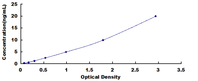
0.312-20ng/mL. The standard curve concentrations used for the ELISA’s were 20ng/mL, 10ng/mL, 5ng/mL, 2.5ng/mL, 1.25ng/mL, 0.625ng/mL, 0.312ng/mL.
SENSITIVITY
The minimum detectable dose of UGGT1 is typically less than 0.113ng/mL.
The sensitivity of this assay, or Lower Limit of Detection (LLD) was defined as the lowest protein concentration that could be differentiated from zero. It was determined by adding two standard deviations to the mean optical density value of twenty zero standard replicates and calculating the corresponding concentration.
SPECIFICITY
This assay has high sensitivity and excellent specificity for detection of UGGT1.
No significant cross-reactivity or interference between UGGT1 and analogues was observed.
You can reference link of the kit as following
https://dldevelop.com/Research-reagent/dl-uggt1-hu.html
https://www.dldevelop.com/uploadfile/data/DL-UGGT1-Hu.pdf
Introduction
| Item | Standard | Test | |
| Description |
The kit is a sandwich enzyme immunoassay for the in vitro quantitative measurement of UGGT1 in human tissue homogenates, cell lysates and other biological fluids. |
Conform | |
| Identification | Colorimetric | Positive | |
| Composition | Traditional ELISA Kit | Ready-to-Use ELISA KIT | Conform |
| Pre-coated, ready to use 96-well strip plate 1 | Pre-coated, ready to use 96-well strip plate 1 | ||
| Plate sealer for 96 wells 2 | Plate sealer for 96 wells 2 | ||
| Standard 2 | Standard 2 | ||
| Diluents buffer 1×45mL | Standard Diluent 1×20mL | ||
| Detection Reagent A 1×120μL | Detection Solution A 1×12mL | ||
| Detection Reagent B 1×120μL | Detection Solution B 1×12mL | ||
| TMB Substrate 1×9mL | TMB Substrate 1×9mL | ||
| Stop Solution 1×6mL | Stop Solution 1×6mL | ||
| Wash Buffer (30 × concentrate) 1×20mL | Wash Buffer (30 × concentrate) 1×20mL | ||
| Instruction manual 1 | Instruction manual 1 | ||
Test principle
The microtiter plate provided in this kit has been pre-coated with an antibody specific to the index. Standards or samples are then added to the appropriate microtiter plate wells with a biotin-conjugated antibody preparation specific to the index. Next, Avidin conjugated to Horseradish Peroxidase (HRP) is added to each microplate well and incubated. After TMB substrate solution is added, only those wells that contain the index, biotin-conjugated antibody and enzyme-conjugated Avidin will exhibit a change in color. The enzyme-substrate reaction is terminated by the addition of sulphuric acid solution and the color change is measured spectrophotometrically at a wavelength of 450nm ± 10nm. The concentration of the index in the samples is then determined by comparing the O.D. of the samples to the standard curve.
Recovery
Matrices listed below were spiked with certain level of recombinant and the recovery rates were calculated by comparing the measured value to the expected amount of the index in samples.
| Matrix | Recovery range (%) | Average(%) |
| serum(n=5) | 81-93 | 86 |
| EDTA plasma(n=5) | 80-97 | 88 |
| heparin plasma(n=5) | 90-101 | 95 |
Linearity
The linearity of the kit was assayed by testing samples spiked with appropriate concentration of the index and their serial dilutions. The results were demonstrated by the percentage of calculated concentration to the expected.
| Sample | 1:2 | 1:4 | 1:8 | 1:16 |
| serum(n=5) | 82-96% | 83-98% | 81-99% | 93-101% |
| EDTA plasma(n=5) | 88-101% | 86-95% | 90-102% | 80-93% |
| heparin plasma(n=5) | 80-91% | 82-90% | 95-104% | 79-95% |
Precision
Intra-assay Precision (Precision within an assay): 3 samples with low, middle and high level the index were tested 20 times on one plate, respectively.
Inter-assay Precision (Precision between assays): 3 samples with low, middle and high level the index were tested on 3 different plates, 8 replicates in each plate.
CV(%) = SD/meanX100
Intra-Assay: CV<10%
Inter-Assay: CV<12%
Stability
The stability of ELISA kit is determined by the loss rate of activity. The loss rate of this kit is less than 5% within the expiration date under appropriate storage conditions.
Note:
To minimize unnecessary influences on the performance, operation procedures and lab conditions, especially room temperature, air humidity and incubator temperatures should be strictly regulated. It is also strongly suggested that the whole assay is performed by the same experimenter from the beginning to the end.
Assay procedure summary
1. Prepare all reagents, samples and standards;
2. Add 100µL standard or sample to each well. Incubate 2 hours at 37℃;
3. Aspirate and add 100µL prepared Detection Reagent A. Incubate 1 hour at 37℃;
4. Aspirate and wash 3 times;
5. Add 100µL prepared Detection Reagent B. Incubate 1 hour at 37℃;
6. Aspirate and wash 5 times;
7. Add 90µL Substrate Solution. Incubate 15-25 minutes at 37℃;
8. Add 50µL Stop Solution. Read at 450nm immediately.
Order or get a Quote
We will reply you within 24 hours!


